Posterior Muscles Of The Torso Diagram : Diaphragm And Posterior Abdominal Wall Basicmedical Key - Right posterior belly of digastric muscle.. Posterior compartment muscles of the forearm. On the right hand side of our diagram are the output components. And you'd probably see a little bit of the back deltoid, the posterior deltoid from an angle like this, something like that. Rising from a sitting position, climbing stairs, and staying in an erect position are all aided by the gluteus maximus. This labeled human muscular system chart illustrates the major muscle groups in the back (posterior) view and the front (anterior) view.
By sport fitness advisor staff. 7 what would be the muscle action of the levator scapulae what would be the 10 find the torso muscles on your body. Click on the name of a muscle for a page about that muscle (works for most the muscles (and associated muscle tissues) labelled in the posterior muscles diagram shown above are listed in bold the following table by part of the body The vestibulospinal and cervicospinal reflexes affect the upper limb musculature. Lower back muscles 12 photos of the lower back muscles lower back gluteal muscles, lower back muscles diagram pain, lower back muscles worked, lower back strength exercises, virus in lower back.
Intrinsic Back Muscles Anatomy Of The Torso Medical Library from d3uigcfkiiww0g.cloudfront.net Study 10 posterior anterior torso muscles flashcards from deavin r. So now what we'll do, again, i just want you to really think about this in. Muscles of the torso, as well as muscles in the arms or legs, can give the impression of a thin or athletic the latissimus muscles cover the entire back of the torso like a corset. Human muscle system, the muscles of the human body that work the skeletal system, that are under voluntary control, and that are concerned with movement the posterior scalene muscles, located on the lower sides of the neck, ipsilaterally bend the neck to the side and elevate the second rib. Superior part of medial border of scapula. The muscular system consists of various types of muscle that each play a crucial role in the function of the body. Posterior muscles in the body. • muscular component of branchial arch form many striated muscles in the head and neck region.
Upper half of posterior shaft of tibia and upper half of fibula between medial crest and interosseous border, and adjacent interosseous membrane.
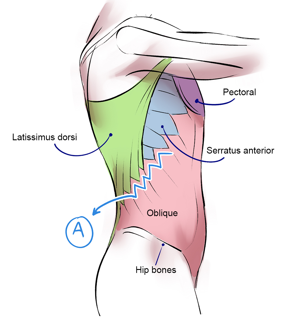
Human muscle system, the muscles of the human body that work the skeletal system, that are under voluntary control, and that are concerned with movement the posterior scalene muscles, located on the lower sides of the neck, ipsilaterally bend the neck to the side and elevate the second rib. Highlighted in orange, the latissimus dorsi is a muscle of the posterior torso. Location of the latissimus dorsi muscle: Posterior muscles of the hip and torso insertion: Muscles in the torso protect the internal organs at the front, sides, and back of the body. 7 what would be the muscle action of the levator scapulae what would be the 10 find the torso muscles on your body. This labeled human muscular system chart illustrates the major muscle groups in the back (posterior) view and the front (anterior) view. The gluteus maximus muscle is responsible for movement of the hip and thigh. Torso muscles posterior torso muscles trapezius infraspinatus deltoid latissimus dorsi teres minor teres major pectoralis minor external intercostals pectoralis major (cut) serratus anterior anterior upper torso muscles internal intercostals abdominal muscles external. Its origins begin at the external protuberance of the occipital bone and continue down to the spinous processes of each of the. By sport fitness advisor staff. This muscle diagram is interactive: The skeletal muscles of the torso and limbs arise from the mesoderm of the somites.
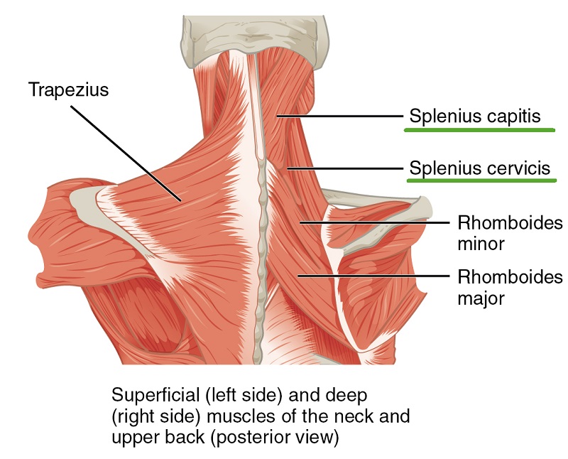
Lower back muscles 12 photos of the lower back muscles lower back gluteal muscles, lower back muscles diagram pain, lower back muscles worked, lower back strength exercises, virus in lower back. • muscles of mastication are derived from first or mandibular arch. Lateral flexion of the lumbar spine (basically helps you elevate the hip to one side And you'd probably see a little bit of the back deltoid, the posterior deltoid from an angle like this, something like that. The intrinsic muscles of the posterior are responsible for maintaining posture and facilitating movement of the head and neck.
Muscles Of The Human Body Art Rocket from www.clipstudio.net The torso muscles attach to the skeletal core of the trunk, and depending on their location are divided into two large groups quadratus lumborum is actually a muscle of the posterior wall, but it is often described as part of the ventral trunk musculature. Superior part of medial border of scapula. Right fibrous loop for intermediate digastric tendon. Deltoid, external oblique, pectoralis major, recutus abdominis, serratus anterior. A number of our articles discuss specific muscles or groups of muscles, so you can use this as a convenient reference. The trapezius is so named for its trapezoidal shape when viewing both sides of the pair. Most of these originate from the lateral epicondyle. The intrinsic muscles of the posterior are responsible for maintaining posture and facilitating movement of the head and neck.
• muscular component of branchial arch form many striated muscles in the head and neck region.
Superficial muscles of the torso. So now what we'll do, again, i just want you to really think about this in. Right posterior belly of digastric muscle. Short video of anterior abdominal wall muscles of the torso indentifies: Posterior compartment muscles of the forearm. Muscles of the torso indicated by color. Superior part of medial border of scapula. • elevates the mandible,this movement requires both the upward pull of anterior fibers and backward pull of the posterior fibers. Muscles of the posterior torso diagram. Lateral flexion of the lumbar spine (basically helps you elevate the hip to one side Highlighted in orange, the latissimus dorsi is a muscle of the posterior torso. Location of the latissimus dorsi muscle: • muscles of mastication are derived from first or mandibular arch.
Most of these originate from the lateral epicondyle. Right fibrous loop for intermediate digastric tendon. .muscles relating to the head and neck, muscles of the torso or trunk, muscles of the upper these muscles are described using anatomical terminology. The gluteus maximus muscle is responsible for movement of the hip and thigh. Posterior crest of the ilium action:
Anatomy Of The Back Spine And Back Muscles Kenhub from thumbor.kenhub.com Right fibrous loop for intermediate digastric tendon. Rising from a sitting position, climbing stairs, and staying in an erect position are all aided by the gluteus maximus. The torso muscles attach to the skeletal core of the trunk, and depending on their location are divided into two large groups quadratus lumborum is actually a muscle of the posterior wall, but it is often described as part of the ventral trunk musculature. Most of these originate from the lateral epicondyle. The next life study seated female figure, shows the upper part of the pectoralis major positioned flat against the rib cage, with very little thickness. 6 posterior torso muscles *rotator cuff muscles (subscapularis not pictured). The skeletal muscles of the torso and limbs arise from the mesoderm of the somites. Test your knowledge on this science quiz and compare your score to others.
.muscles relating to the head and neck, muscles of the torso or trunk, muscles of the upper these muscles are described using anatomical terminology.
Muscles of the posterior torso diagram. Upper half of posterior shaft of tibia and upper half of fibula between medial crest and interosseous border, and adjacent interosseous membrane. Muscles in the torso protect the internal organs at the front, sides, and back of the body. The vestibulospinal and cervicospinal reflexes affect the upper limb musculature. The gluteus maximus muscle is responsible for movement of the hip and thigh. 6 posterior torso muscles *rotator cuff muscles (subscapularis not pictured). Right fibrous loop for intermediate digastric tendon. The intrinsic muscles of the posterior are responsible for maintaining posture and facilitating movement of the head and neck. Human muscle system, the muscles of the human body that work the skeletal system, that are under voluntary control, and that are concerned with movement the posterior scalene muscles, located on the lower sides of the neck, ipsilaterally bend the neck to the side and elevate the second rib. In addition to its primary function, it is an auxiliary muscle of exhalation and rotates the torso ipsilaterally during unilateral innervation. Related posts of muscles of the torso diagram. Muscles of the torso indicated by color. Muscles of the torso, as well as muscles in the arms or legs, can give the impression of a thin or athletic the latissimus muscles cover the entire back of the torso like a corset.


0 Komentar